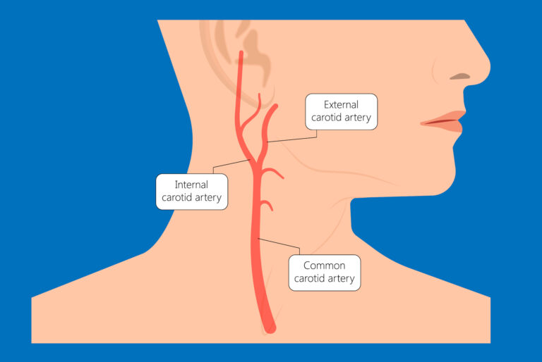
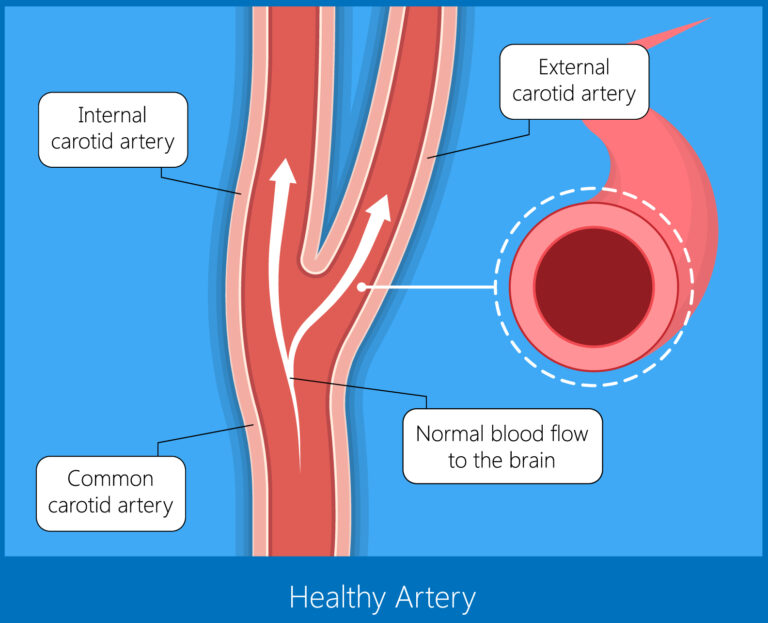
The Carotid artery is a blood vessel supplying the brain and face. It is a paired artery in the neck – one on the right and other on the left. The internal carotid artery is a branch of the common carotid artery which supplies the brain. The external carotid artery supplies the face.
2. What is Carotid artery disease (or Carotid artery stenosis)?
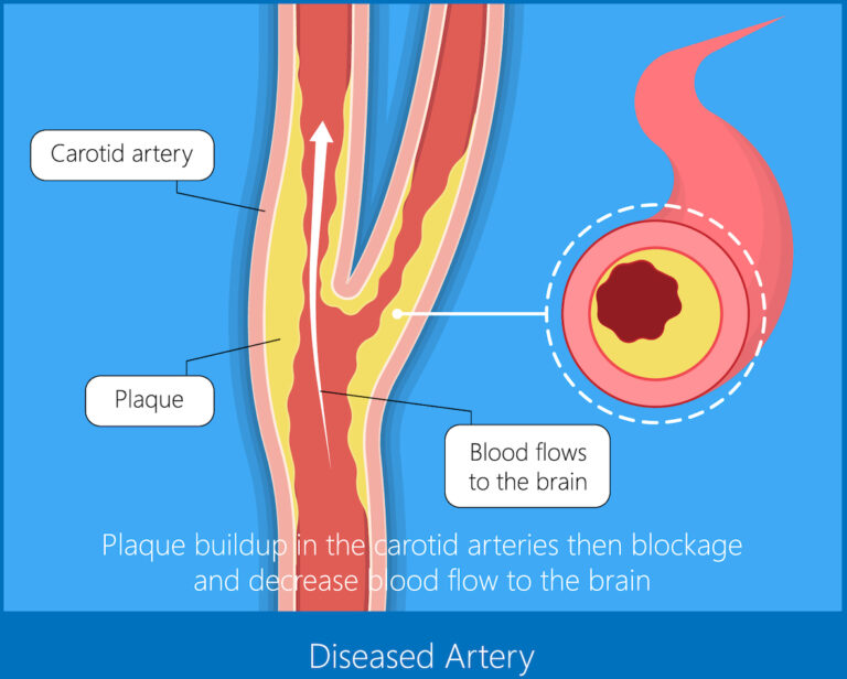
Narrowing of the Carotid artery is referred as Carotid artery disease (or Carotid artery stenosis). The narrowing occurs due to a slow progressive process called Atherosclerosis. In this process, there is deposition of a material called ‘Atherosclerotic plaque’ in the wall of the artery. This plaque is made of fat, cholesterol, calcium and other substances.
3. What are the symptoms of Carotid artery disease?
‘Patients with Carotid artery stenosis present with Stroke or a ‘mini-stroke’ (called ‘Transient ischemic attack’ or TIA). In stroke, the symptoms are permanent, whereas, in mini-stroke, the symptoms are temporary.
The stroke (or mini-stroke/TIA) symptoms include
Inability to use one side of the body
Disturbances in speech
Loss of sensation of one side of the body
Disturbances in vision
Carotid artery stenosis can also progress without any symptoms in some patients
4. What are the causes for carotid artery disease?
There are multiple factors which contribute to the development of Carotid artery disease.
Diabetes
Smoking
Hypertension (High BP)
Increased blood cholesterol
Obesity
Kidney disease (‘Chronic kidney disease’)
Family history
5. How is Carotid artery disease diagnosed?
The following tests help in detecting a Carotid artery stenosis
Carotid doppler ultrasound – This is a screening test. It is usually the first test done after Carotid artery disease is suspected. This test is also useful for screening in patients without any symptoms. No radiation is involved in this study.
CT angiography– This test gives more detailed images about the carotid arteries compared to Carotid doppler ultrasound. This test also shows the brain and the arteries of the brain simultaneously. CT scan uses radiation to obtain the images of the carotid and brain arteries.
MR angiography – This test also gives information about the carotid and brain arteries. It also gives better images of the brain compared to a CT scan. Also, this study does not involve radiation exposure. However, the level of detail in the images of the carotid and brain arteries is inferior compared to CT angiography
Digital subtraction angiography (DSA) – This test is considered the best tool (gold-standard test)to study the carotid and brain arteries. This is an invasive study unlike the 3 studies described above. The test involves a physician advancing a small tube (called catheter) through the groin (femoral artery) or through the wrist (radial artery). The catheter is advanced using “Live X-ray” (fluoroscopic) guidance into the carotid arteries and vertebral arteries. Images are obtained by injecting iodinated contrast.
6. What are the treatment options for carotid artery disease?
Taking medicines as prescribed – Anti-platelet medications (eg. Aspirin, Clopidogrel, Ticagrelor etc) and lipid lower medications (‘Statins’) are routinely prescribed medicines. In addition, medications to adequately control BP and blood sugar are also of utmost importance.
Procedures to improve flow of the affected Carotid artery – Some patients, in addition to the lifestyle changes and medications, will need additional procedures to prevent a life-threatening future stroke. The procedures include surgical (Carotid Endarterectomy) and Interventional (Carotid artery stenting)
7. What is Carotid artery stenting? How does it treat the disease?
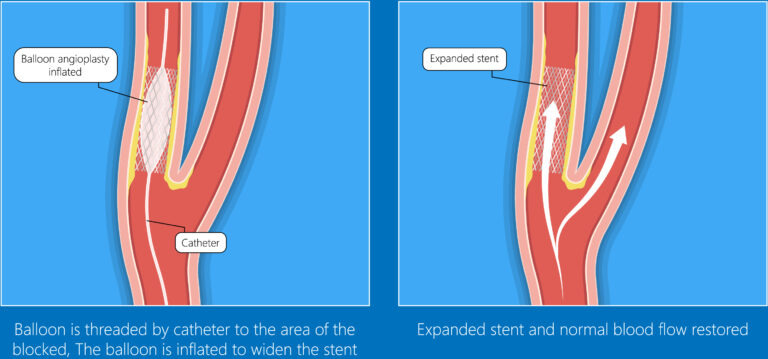
Carotid artery stenting is performed by placing a small mesh called stent across the narrowed segment of the carotid. The stent pushes the ‘atherosclerotic plaque’ away, thereby eliminating the restriction to the blood flow.
The procedure is performed under local anaesthesia. Just like an angiogram, a small tube called a catheter is advanced through the groin (femoral artery) or the wrist (radial artery) and advanced into the carotid artery. Through these tubes, fine wires are advanced across the carotid artery stenosis. The stenosis is dilated using a balloon. Once the pathway is widened with the balloon, the stent is then placed which completes the treatment
8. Can we prevent Carotid artery disease? What are the lifestyle changes that are needed?